Figures & data
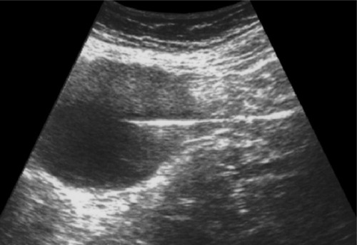
Figure 1 Ultrasonographic view of the cyst adherent to the uterine fundus.
Note: Myometrial calcifications are visible (small arrows).
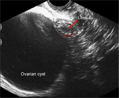
Figure 2 Ultrasonographic guidance of the puncture.
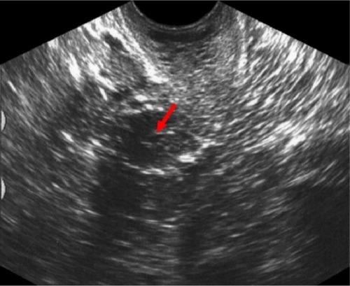
Figure 3 Ultrasound scan after withdrawal of the needle.
Note: Small amount of the liquid in the uterine cavity after hysteroscopy (arrow).