Figures & data
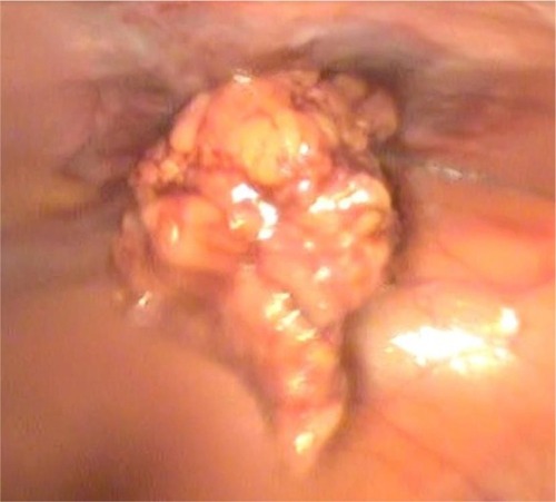
Figure 1 (A, B) Ectopic localization of the IUD by X-ray imaging.
Abbreviation: IUD, intrauterine device.
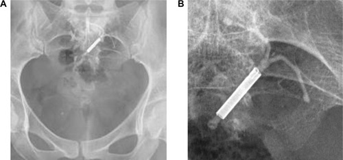
Figure 2 Ultrasound imaging of the IUD.
Notes: (A) Ectopic localization of the IUD in ultrasound imaging (8 mm from the left common iliac artery). (B) Ectopic localization of the IUD in ultrasound imaging. The hypoechoic halo is visible. (C) Ectopic localization of the IUD in ultrasound imaging: increased vascularity around the foreign body is visible, particularly around the IUD ends.
Abbreviation: IUD, intrauterine device.
Abbreviation: IUD, intrauterine device.
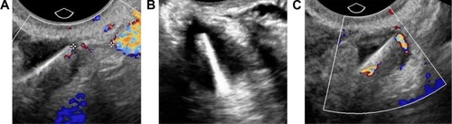
Figure 3 A piece of the IUD string (pointed with arrows) visible on the omental surface.
Abbreviation: IUD, intrauterine device.
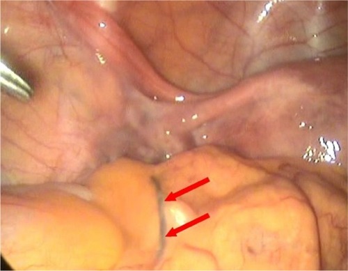
Figure 4 IUD embedded in the omentum.
Note: Encapsulation of the vertical arm of the IUD (pointed with arrow).
Abbreviation: IUD, intrauterine device.
Abbreviation: IUD, intrauterine device.
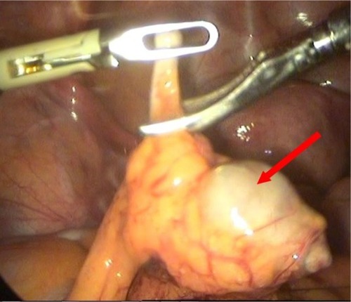
Figure 5 Bipolar electroresection of the part of the omentum with the ingrown IUD.
Abbreviation: IUD, intrauterine device.
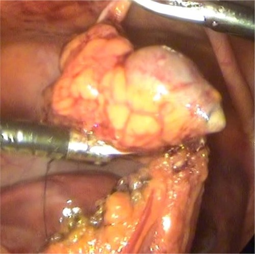
Figure 6 Removal of the IUD from the abdominal cavity.