Figures & data
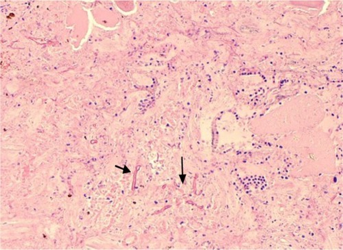
Figure 1 Left upper lobe round infiltrate in chest X-ray.
Note: The arrow indicate the cavitary lesion in the lung.
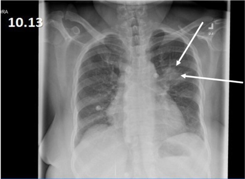
Figure 2 Computed tomography scan of chest showing left upper lobe lung lesion with cavitations, along with diffuse bilateral ground glass opacification.
Notes: The arrow indicate the cavitary lesion in the lung. The inset shows that the cavitary lesion in lung is on that level on coronal view.
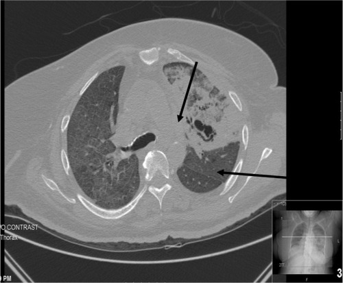
Figure 3 Broad-based pauci-septate hyphae with dichotomous wide-angle branching (arrow) on microscopic examination of Lung.
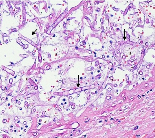
Figure 4 Broad-based pauci-septate hyphae with dichotomous wide-angle branching (arrow) on microscopic examination of thyroid.