Figures & data
Figure 1 Fundus photographs showing veins are convoluted and slightly dilated, but arteries have straight courses.
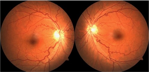
Figure 2 The cilioretinal artery course inferotemporally from behind the disc margin (arrow).
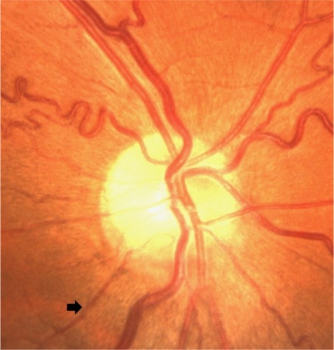
Figure 3 The cilioretinal artery begins to fluoresce at early phase angiogram (13.7 seconds).
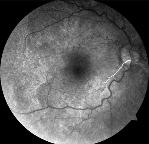
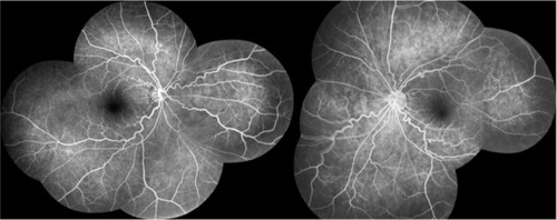
Figure 4 FFA photography of both eyes showing obviously tortuous and dilated retinal veins.
Note: No leakage or evidence of dye is seen.
Abbreviation: FFA, fluorescein fundus angiography.
Abbreviation: FFA, fluorescein fundus angiography.