Figures & data
Figure 1 Slit lamp microscopic images of the left eye at the time of the initial presentation.
Notes: (A) A corneal ulcer with corneal opacity on the lower part of the cornea and conjunctival injection appeared. (B) Slit lamp image of the cornea with fluorescein staining showed defect of corneal epithelium. Chemosis and/or conjunctivochalasis on the lower part of burbar conjunctiva were shown.
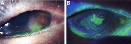
Figure 2 Slit lamp images from 2 months after the start of treatments.
Notes: (A) Despite the treatment, the corneal epithelial defect and opacity on the lower part of the cornea persisted. (B) Slit lamp image with fluorescein staining showed that the area of the corneal epithelium defect reduced. Chemosis and/or conjunctivochalasis were still shown.
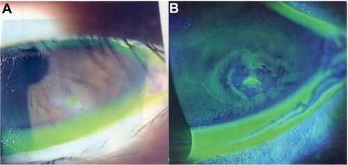
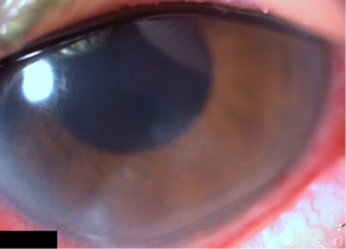
Figure 3 Slit lamp image from 3 weeks after the start of treatments with Mucosta ophthalmic suspension.
Notes: The corneal epithelial defect had then resolved, even though subepithelial opacity slightly remained. As this picture was taken after vitrectomy for treatment of vitreous hemorrhage, conjunctival hyperemia was shown.