Figures & data
Figure 1 Interval fundus photography.
Notes: Color fundus photographs at presentation (A), 2 months (B), 3 months (C), and 12 months (D). At month 3, there is no clinical evidence of leukemic infiltration in the right eye (hemorrhages only) with a small residual area remaining just below the foveal center in the left. All lesions and intraretinal hemorrhages in the macula resolved by month 12.
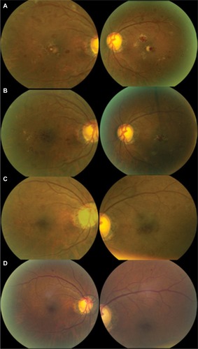
Figure 2 Serial spectral domain OCT images, with image registration, through the fovea at presentation (A), 2 months (B), 3 months (C), and 12 months (D).
Notes: (A) Initially, there were large inner foveal hyperreflective lesions in both eyes with a second outer retinal lesion in the right. (B) At 2 months, there were numerous smaller foci in the inner retina present after the larger lesion regressed in both eyes. There was a new area of outer retinal involvement in the left fovea. (C) There is complete resolution of the leukemic infiltrates in the right eye with minimal inner retinal involvement at the fovea in the left. (D) At 12 months, there was complete restoration of the foveal contour with focal residual disruption of the photoreceptor outer segments in each eye.
Abbreviations: CMT, central macular thickness; Mac Vol, macular volume; OCT, optical coherence tomography; OD, oculus dexter; OS, oculus sinister.
Abbreviations: CMT, central macular thickness; Mac Vol, macular volume; OCT, optical coherence tomography; OD, oculus dexter; OS, oculus sinister.
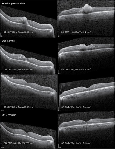
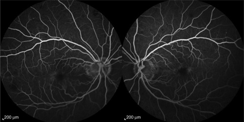
Figure 3 Intravenous fluorescein angiography at month 3.
Notes: There is no evidence of retinal vascular occlusion with minimal late perifoveal leakage. The foveal avascular zone and perifoveal capillary network are preserved. Images were obtained using a 55 degree objective.