Figures & data
Figure 1 Gangrenous digits of the patient.
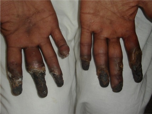
Figure 2 Ulcerated jugulodigastric node.
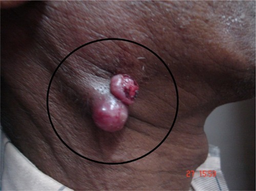
Figure 3 Diagnostic flowchart for Raynaud’s Phenomenon.
Notes: * Refers to polyneuropathy, organomegaly, endocrinopathy, monoclonal gammopathy, and Skin changes. It is a rare multisystemic disease that occurs in the setting of a plasma cell dyscrasia.
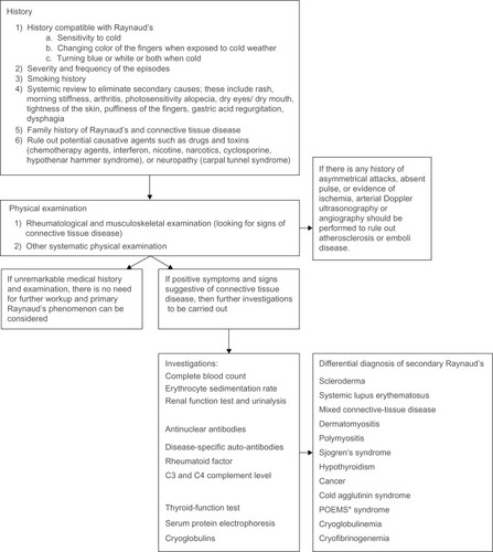
Table 1 Routine blood investigations
Table 2 Immunology screening
Table 3 Immunoglobulin screening
Table 4 Virology screening
Table 5 Other tests
Table 6 Imaging studies
Figure 4 Histological specimen of jugulodigastric lymph node
Notes: (A) Biopsy from the ulcerated jugulodigastric node showing spindle cell carcinoma. (B) Cytology smear from the ulcerated jugulodigastric node showing spindle cell carcinoma.
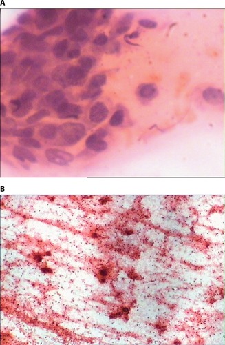
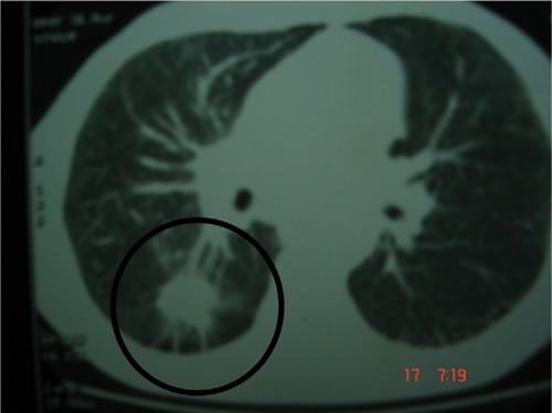
Figure 5 Lymphangitis carcinomatosis involving the right lobe and mediastinal adenopathy.