Figures & data
Figure 1 Dense vitritis.
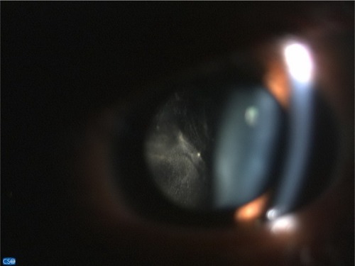
Figure 2 Intraoperative findings were dense vitritis, fibrous tissue on superior disk with diffuse periphery sclerotic vessels, and sheathing of both artery and vein.
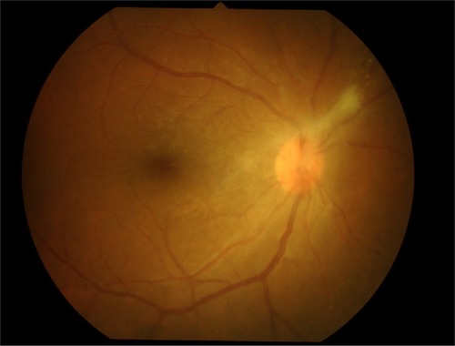
Figure 3 Fluorescein angiography.
Notes: (A) Photo fundus showed fibrous band on hyperemia optic disk. (B) Optic disk showed leakage in early phase. (C–D) Ischemic area was observed at periphery with occluded retinal artery and leakage retinal vein in all quadrants of the right eye.
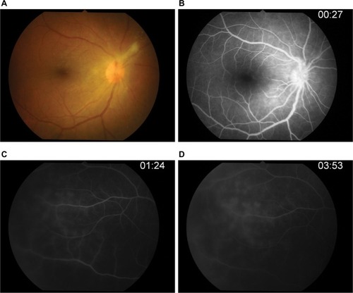
Figure 4 Panretinal photocoagulation of the right eye at ischemic area was performed.
Notes: (A) Posterior pole. (B) Laser scar at periphery.
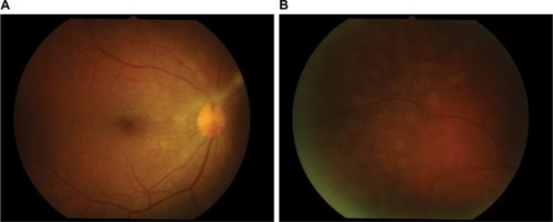
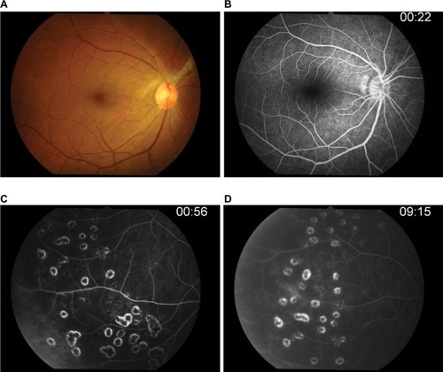
Figure 5 Post-IV acyclovir after 14 days of therapy.
Notes: (A) Laser scar. (B) Repeated FA, peripheral retinal vein still has leakage. (C–D) Late phase.
Abbreviation: IV, intravenous; FA, fluorescein angiography.
Abbreviation: IV, intravenous; FA, fluorescein angiography.
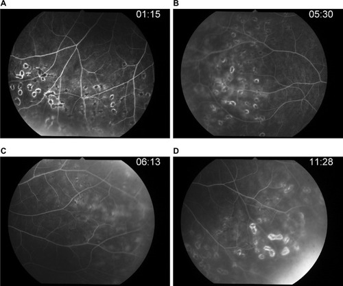
Figure 6 Post-IV acyclovir after 4 weeks of therapy.
Notes: (A) Photo fundus showed optic disk edema decreased. (B–D) FA confirmed retinal phlebitis improved.
Abbreviation: IV, intravenous; FA, fluorescein angiography.
Abbreviation: IV, intravenous; FA, fluorescein angiography.
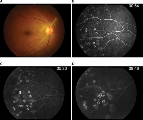
Figure 7 Post-acyclovir after 3 months of therapy.
Notes: (A) Normal optic disk. (B–D) Retinal vascular leakage was not observed.
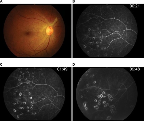
Figure 8 Post-acyclovir after 6 months of therapy (after 3 months of discontinued oral acyclovir).
Notes: (A) There was no vitreous haze and disk edema. (B) Optic disk showed no leakage. (C–D) Periphery has become quiet.