Figures & data
Figure 1 Clinical picture of corneal endothelial graft rejection at baseline.
Note: Slit lamp biomicroscopy with anterior segment photography at a magnification of ×16 shows intense conjunctival hyperemia and severe corneal edema with diffuse Descemet’s folds.
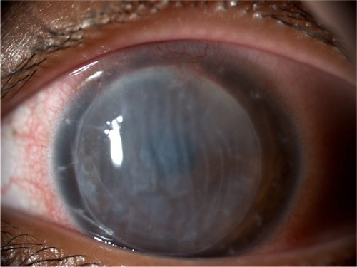
Figure 2 Confocal microscopy image at baseline.
Note: Confocal microscopy image shows increased number of dendritic cells.
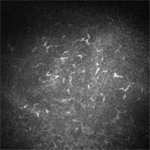
Figure 3 Clinical picture of corneal endothelial graft rejection 1 month after the injection.
Note: Slit lamp biomicroscopy with anterior segment photography at a magnification of ×16 shows conjunctival hyperemia recovery and improvement in corneal transparency.
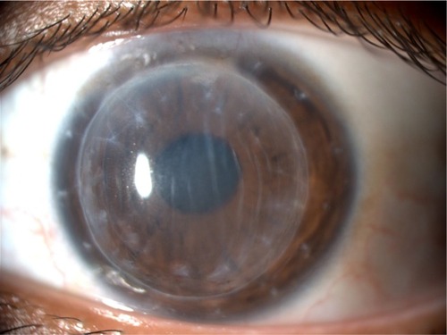
Figure 4 Clinical picture of corneal endothelial graft rejection 3 months after the injection.
Note: Slit lamp biomicroscopy with anterior segment photography at a magnification of ×16 shows full recovery of corneal transparency with few residual Descemet’s folds not involving visual axis.
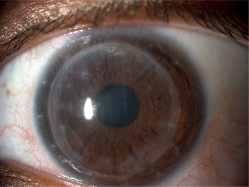
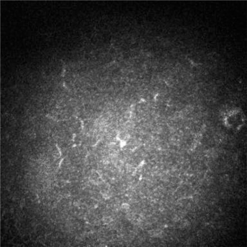
Figure 5 Confocal microscopy image 3 months after the injection.
Note: Confocal microscopy image shows dendritic cell number within the physiological range.