Figures & data
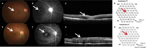
Figure 1 Evaluation of anatomical and functional status before and after treatment using FA, OCT, and ERG.
Notes: (A) Ischemic retinopathy with alteration of foveal reflex (arrow) in the right eye. (B) Retinal FA showing ischemic retinopathy, especially involving the macular area (arrow). (C) OCT showed thickening and hyperreflectivity of the inner retinal layers (arrow). (D) Multifocal ERG showed a decrease in electrical responses from central macula (arrow). (E) Resolution of cotton wool spots (arrow). (F) Reperfusion of some previously ischemic areas during examination with fluorescein (arrow). (G) OCT showed formation of epiretinal membrane (arrow). (H) Multifocal ERG remained at low amplitudes corresponding to central area (arrow), indicating macular dysfunction.