Figures & data
Figure 1 Color fundus image of the left eye showed an area of retina whitening, extending along the distribution of three cilioretinal arteries.
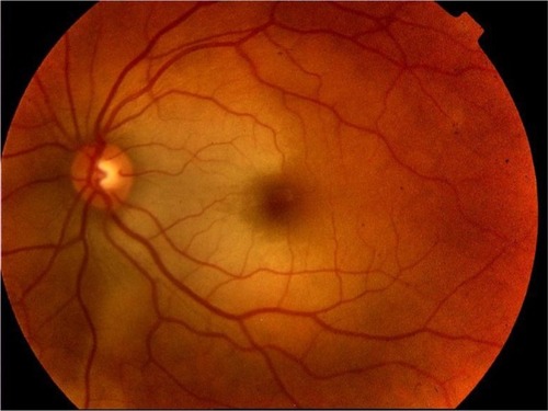
Figure 2 Fluorescein angiography images revealed only a vertical choroidal watershed centered on the optic disk, a faint screen effect in the perifoveal area due to intraretinal edema, and a normal CRA perfusion.
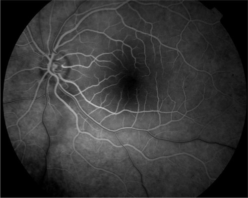
Figure 3 A horizontal, cross-sectional OCT tomogram through the fovea disclosed diffuse thickening of the neurosensory retina.
Abbreviations: OCT, optical coherence tomography; N, nasal; S, superior; T, temporal; I, inferior.
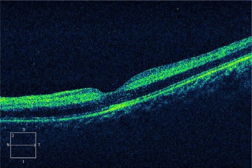
Figure 4 Visual field of the left eye (SITA-Fast Central 30-2 strategy) immediately (A) and after 3 months (B) since CRA occlusion.
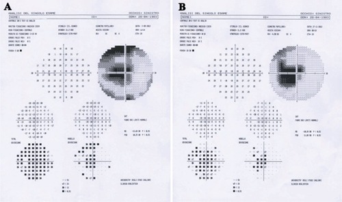
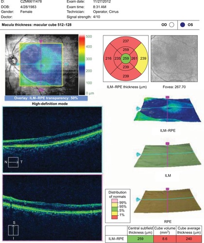
Figure 5 The macular thickness map, which was generated by means of OCT after 3 months since CRA occlusion, demonstrated thinning along the distribution of three cilioretinal arteries.