Figures & data
Figure 1 Initial ocular manifestations.
Notes: (A) Slit-lamp photographs showed anterior chamber inflammation and severe conjunctivitis in both eyes. Fluorescein leakage was observed in the anterior chamber. (B) Fundus photographs showed mild optic disk edema and disk hemorrhage in the left eye. (C) Fluorescein angiographic images showed dye leakage at the optic disk in the left eye. (D) Optical coherence tomographic (OCT) images showed no abnormal foveal findings in both eyes.
Abbreviations: R, right; L, left.
Abbreviations: R, right; L, left.
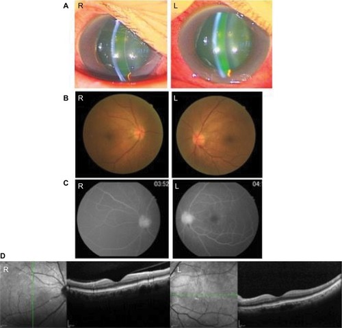
Figure 2 Visual field tests using a Goldmann perimeter.
Notes: (A) Central and paracentral scotomas were observed in the left eye at the initial ocular examination. (B) Regression of visual field defect was observed in the left eye at 2-month follow-up after pulse steroid therapy.
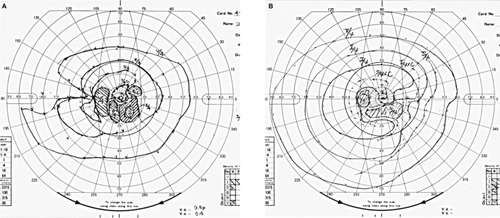
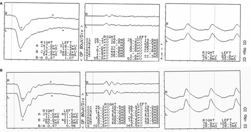
Figure 3 Full-field electroretinograms showing changes before and after pulse steroid therapy.
Notes: (A) At the initial ocular examination, reductions in amplitude of oscillatory potentials and b-wave were observed in both eyes. (B) After pulsed-dose steroid therapy, recovery in oscillatory potentials was observed.