Figures & data
Figure 1 Spectral domain coherence tomography identifies symmetric retinal nerve fiber layer and ganglion layer loss in the left eye (arrow).
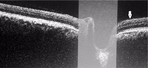
Figure 2 Spectral domain coherence tomography identifies symmetric retinal nerve fiber layer and ganglion layer loss in the right eye (arrow).
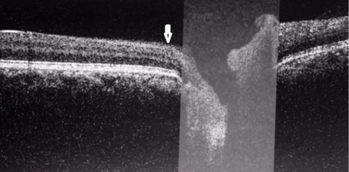
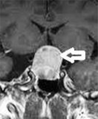
Figure 3 Coronal view, post-contrast T1 brain magnetic resonance imaging, showing a pituitary macroadenoma with compression of the optic chiasm (arrow).