
Figures & data
Figure 1 At presentation, there was linear thickening of skin of lower limbs with small discrete papules and areas of hyperpigmentation.
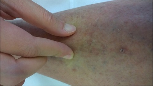
Figure 2 Magnetic resonance imaging of pelvis and legs showed bilateral lower limb subcutaneous edema with prominence of the dermal thickness and patchy tethering with increased T2 signal of the quadriceps and adductor muscle groups.
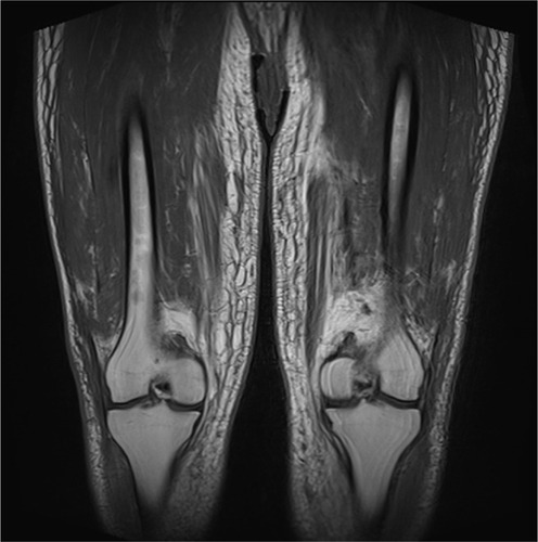
Figure 3 (A) Skin biopsy – leg (×40 magnification), (B) skin biopsy – abdomen (×40 magnification), (C) deep skin biopsy – leg (×100 magnification), and (D) perivascular and thickened collagen – abdomen (×200 magnification).
Note: The histology shows normal epidermis with proliferation of angular fibroblasts in the reticular dermis accompanied by disorganized collagen fibres (A–C). Increased interstitial connective tissue mucin in the dermis associated with perivascular lymphocytic infiltrate, there was thickening of the dermal collagen bundles with loss of adipose tissues around the eccrine glands (D).
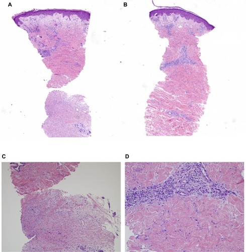
Figure 4 Progress at 9 months: spontaneous regression of skin changes.
