Figures & data
Figure 1 Lipemic serum after centrifugation.
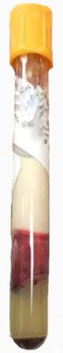
Figure 2 Lipemia retinalis before and after treatment. Fundus examination disclosed bilateral whitish optic discs, diffuse creamy retinal vessels in the peripheral and posterior pole OD (A), OS (B), and salmon-pink retina that were identified as signs of severe lipemia retinalis before treatment. Fundus examination depicted complete bilateral resolution of lipemic retinal vessel disease OD (C), OS (D) after treatment.
Abbreviations: OD, oculus dexter; OS, oculus sinister.
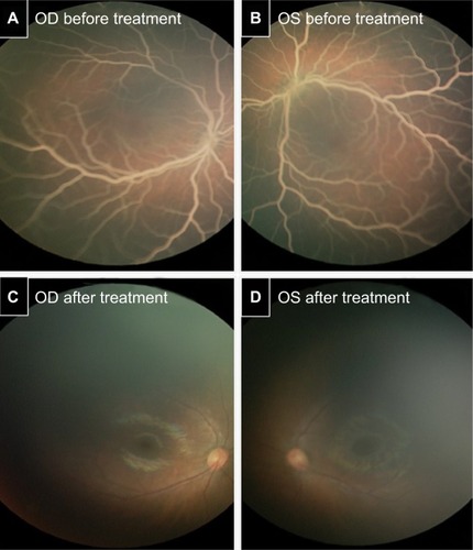
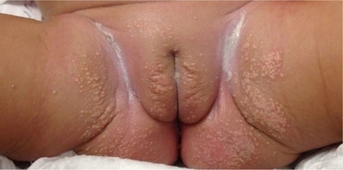
Figure 3 Eruptive vulvar xanthomas.