
Figures & data
Figure 1 A late-onset corneal flap displacement with superior and inferior portion of flap being folded is shown. Bare stroma is exposed and intact nasal hinge is visible under surgical microscope.
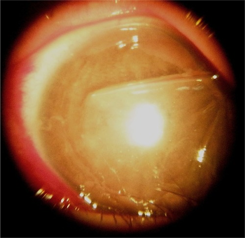
Figure 2 After reposition of the corneal flap a bandage soft contact lens is placed on the cornea for protection under surgical microscope. without epithelial ingrowth.
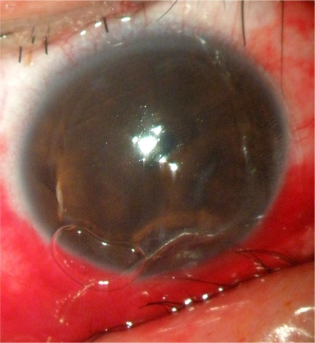
Figure 3 Four months after surgery, the corneal flap is clear and smooth in situ without epithelial ingrowth.
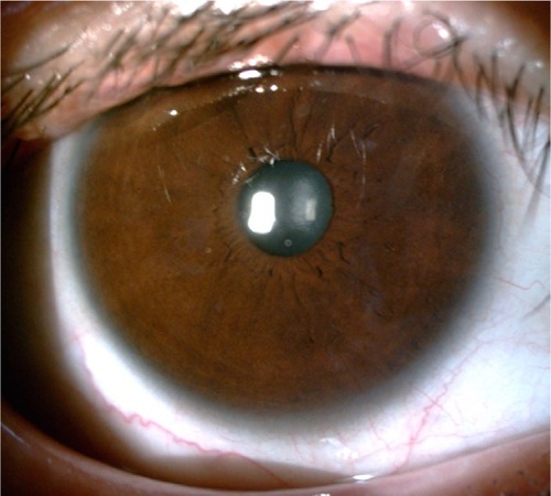
Table 1 Summary of previous literature reporting late flap dislocations or displacements after LASIK
Table 2 Vision prognosis and average age according to mechanisms of injury
Table 3 Percentage of vision after trauma and after treatment according to OTS
Table 4 Percentage of cases with respect to corneal flap conditions after trauma
Table 5 Percentage of cases which received treatments after trauma