Figures & data
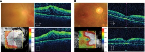
Figure 1 Comparison of fundus and spectral-domain optical coherence tomography images before and after intravenous injection of ranimizumab (IVR) as an adjunct to anti-TB therapy.
Notes: (A) Fundus findings and macula edema associated with CRVO were observed before IVR and anti-TB therapy. (B) CRVO and macular edema were significantly improved after two sessions of IVR.
Abbreviations: CRVO, central retinal vascular occlusion; IVR, injection of ranimizumab; TB, tuberculosis.
Abbreviations: CRVO, central retinal vascular occlusion; IVR, injection of ranimizumab; TB, tuberculosis.
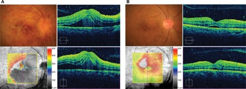
Figure 2 Comparison of fundus and spectral-domain optical coherence tomography images before and after vitrectomy with retinal panretinal photocoagulation.
Notes: (A) Before vitrectomy, retinal hemorrhage in the nasal inferior side with vitreous hemorrhage and relapse of macular edema was detected. (B) After two sessions of vitrectomy with panretinal photocoagulation, retinal hemorrhage and macular edema were resolved, but damage of retinal outer layers in the macula was present.