Figures & data
Figure 1 Anterior segment photograph of the left eye.
Note: Severe inflammation of the anterior chamber and entire anterior synechiae.
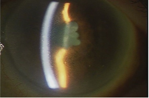
Figure 2 B-scan ultrasonography of the left eye.
Note: The retina was thought to be not detached, and high-intensity opacities of the vitreous body were also absent.
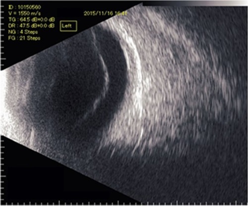
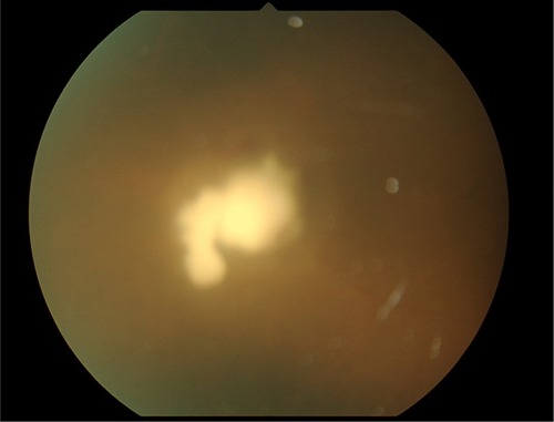
Figure 3 String of pearls OCV in right eye.
Note: OCV was seen continuously from the optic disc.
Abbreviation: OCV, opacitas corporis vitrei.
Abbreviation: OCV, opacitas corporis vitrei.
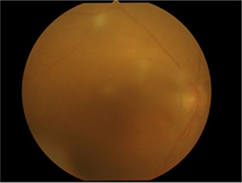
Figure 4 Fungal mass on the right retina.
Notes: A xanthochromistic fungal mass developed on the right temporal retina during systemic therapy. The photo was hazy due to OCV.
Abbreviation: OCV, opacitas corporis vitrei.
Abbreviation: OCV, opacitas corporis vitrei.