Figures & data
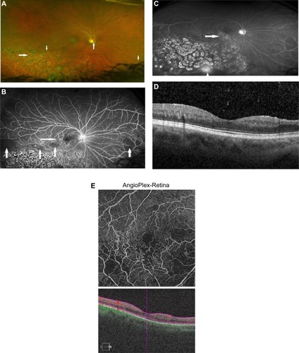
Figure 1 BRVO, left eye.
Notes: (A) Fundus photography demonstrated the site of the occlusion (horizontal arrow) and collateral vessels (vertical arrow). (B) Humphrey visual field 24-2 testing demonstrated an inferior arcuate defect encroaching fixation (arrow). (C) SD-OCT map demonstrated mild thickening centrally and atrophy superotemporally. (D) SD-OCT demonstrated a central macular cyst (larger vertical arrow) and temporal thinning (smaller vertical arrow). (E) OCT-A retina slab (6×6 mm) demonstrated profound capillary nonperfusion in the superotemporal quadrant, involving the foveal avascular zone.
Abbreviations: BRVO, branch retinal vein occlusion; OCT-A, optical coherence tomography-angiography; SD-OCT, spectral domain optical coherence tomography.
Abbreviations: BRVO, branch retinal vein occlusion; OCT-A, optical coherence tomography-angiography; SD-OCT, spectral domain optical coherence tomography.
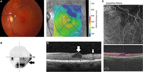
Figure 2 Hemiretinal vein occlusion, right eye.
Notes: (A) Fundus photography demonstrated the site of the occlusion at the disc (upward vertical arrow) and sclerotic veins in the inferotemporal and inferonasal quadrants (downward vertical arrows). Collateral vessels temporal to the center of the macula did not photograph well. Photocoagulation burns (horizontal arrow) are evident. (B) Fluorescein angiography demonstrated areas of capillary nonperfusion temporally and inferonasally (vertical arrows). Collateral vessels temporal to the macula were noted (horizontal arrow). (C) Fluorescein angiography late frames demonstrated mild macular leakage (horizontal arrow) and more prominent leakage inferiorly (vertical arrow). (D) SD-OCT demonstrated no frank cystoid macular edema. (E) OCT-A retina slab (6×6 mm) demonstrated prominent collateral vessels temporal to (and some nasal to) the center of the macula, as well as areas of capillary nonperfusion inferior to the fovea and inferotemporal to the fovea.
Abbreviations: OCT-A, optical coherence tomography-angiography; SD-OCT, spectral domain optical coherence tomography.
Abbreviations: OCT-A, optical coherence tomography-angiography; SD-OCT, spectral domain optical coherence tomography.