Figures & data
Figure 1 Pathology test indicated lung metastasis of renal clear cell carcinoma.
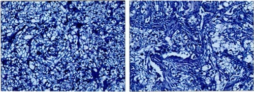
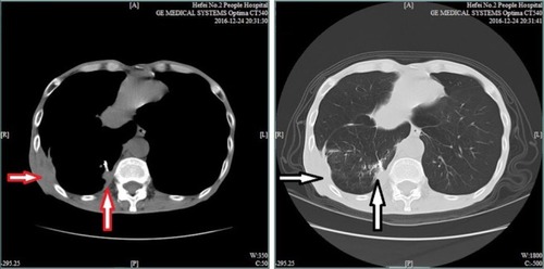
Figure 2 Chest CT scan indicates a large lesion with dimensions of 3.6 cm × 6.3 cm (red and white vertical arrows) located within the basal segment of the lower lobe and extending out of the thoracic cavity, partially complicated with osteolytic destruction (red and white horizontal arrows).
Abbreviation: CT, computed tomography.
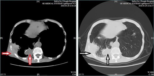
Figure 3 Chest CT scan indicates a larger lesion with dimensions of 2.7 cm × 1.8 cm (red and white vertical arrows) located within the basal segment of the lower lobe and extending out of the thoracic cavity, partially complicated with worse osteolytic destruction (red and white horizontal arrows).
Abbreviation: CT, computed tomography.
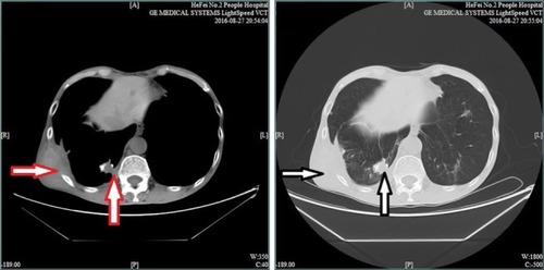
Figure 4 Chest CT scan indicates a larger lesion with dimensions of 2.7 cm × 1.8 cm (red and white vertical arrows) located within the basal segment of the lower lobe and partially extending out of the thoracic cavity, complicated with improved osteolytic destruction (red and white horizontal arrows).
Abbreviation: CT, computed tomography.