Figures & data
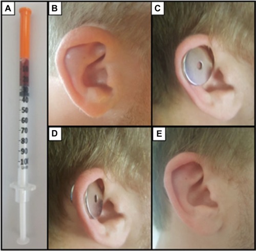
Figure 1 (A) A total of 0.25 mL aspirate using 30G insulin syringe; (B) hematoma can be seen in the scaphoid region cancelling the delineation of the upper crus of the antihelix; (C) metal disc used to reduce the pressure exerted on the auricle; (D) metal disc (anterior) and magnet (posterior) in situ 3-day period; (E) 1-month postdrainage of hematoma with the crus in antihelix clearly seen.