Figures & data
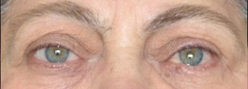
Figure 1 The patient’s appearance on admission to Endocrinology research centre (Moscow, Russia).
Notes: OU (both eyes): exophthalmos; OS (left eye): interpalpebral adhesion, lagophthalmos, hypotropia, and corneal ulcer.
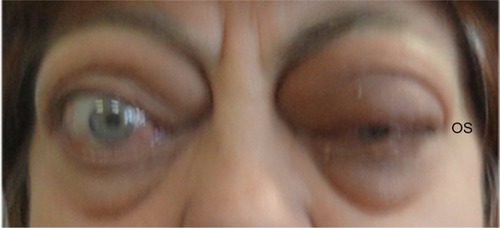
Figure 2 Multislice CT scan (coronal plane) prior to orbital decompression.
Note: There is significant enlargement of the extraocular muscles, more pronounced on the left, and apical crowding.
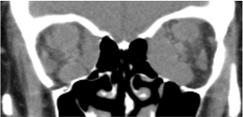
Figure 3 Multislice scan (coronal plane) 1 week after bony decompression of the left orbit.
Notes: There are defects in the lateral, medial, and inferior orbital walls. Significantly enlarged medial and inferior rectus muscles are displaced toward the defects in the corresponding walls. The lateral rectus muscle and orbital fat are observed outside the internal contour of the lateral wall.
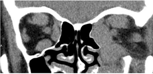
Figure 4 The patient’s appearance 3 months after the orbital decompression and 1 month after the radiation therapy.
Notes: OS (left eye): hypotropia, upper eyelid retraction, lagophthalmos, and superficial keratopathy.
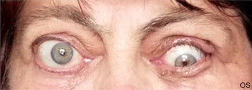
Figure 5 The patient’s appearance 16 months after bony orbital decompression and strabismus surgery. Orthotropy in primary gaze position.