Figures & data
Figure 1 Photographic image of the single-piece trifocal IOL (Pod F FINE VISION).
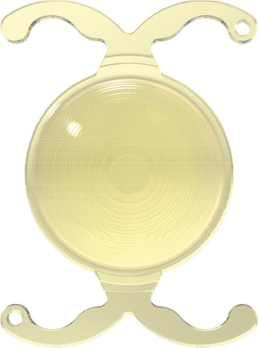
Figure 2 Intraoperative image taken during femtosecond laser surgery.
Notes: (A) 5.0 mm diameter CCC centered on White-to-White (pink circle). (B) Nuclear fragmentation using the cylinder and chop mode (yellow circle and eight pattern).
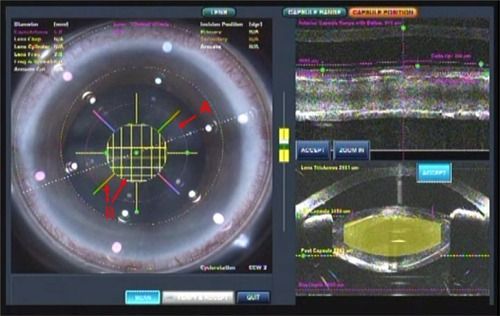
Figure 3 Image of the development of the posterior capsule foramen at the 9 o’clock position (red arrow).
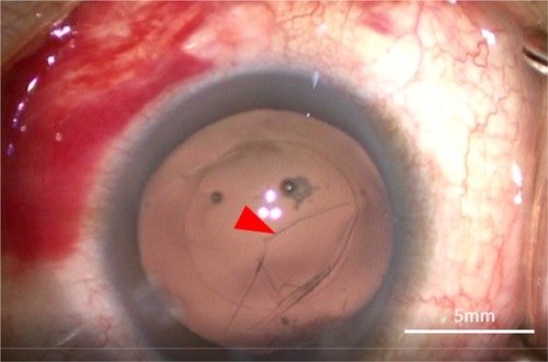
Figure 4 Photographic image of the three-piece +4.0 D bifocal IOL (ZMA00).
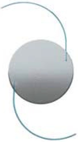
Figure 5 Image of optic capture involving the three-piece +4.0 D bifocal IOL.
Abbreviations: CCC, curvilinear capsulorhexis; IOL, intraocular lens.
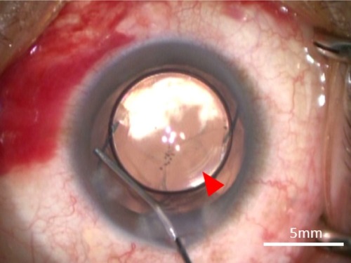
Figure 6 (A) Defocus curve of the Fine Vision trifocal IOL (navy) ZMA00 bifocal IOL (green); right eye with the trifocal IOL (red) and left eye with the bifocal IOL (brown). The defocus curve for the Fine Vision trifocal IOL (navy) and ZMA00 bifocal IOL (green) was provided by the manufacturers. (B) Defocus curve for the right eye with the Fine Vision trifocal IOL (red); left eye with the ZMA00 bifocal IOL (brown) and both eyes (light blue).
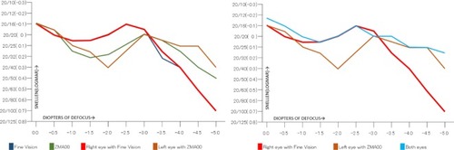
Figure 7 Postoperative contrast sensitivity in both eyes.
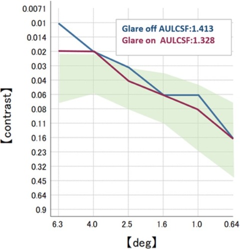
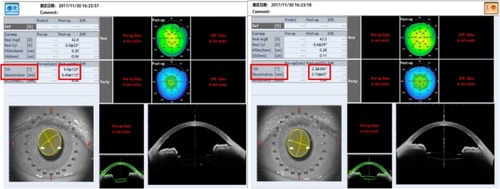
Figure 8 Image of the anterior segment OCT.
Abbreviations: IOL, intraocular lens; OCT, optical coherence tomography.