Figures & data
Figure 1 (A, B) Magnetic resonance imaging scan of abdomen revealed a large well-defined suprarenal mass that measured 12×10×8.3 cm displacing the right kidney inferiorly and inferior the right lobe of liver and close to the porta hepatis, with evidence of cystic changes, fatty component, and calcification.
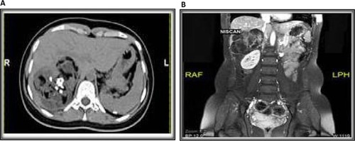
Figure 2 Histopathology of the initial left ovarian tumor demonstrating immature teratoma composed of mixed immature and mature elements including (A) primitive neuroepithelial elements with rosettes formation (H&E, 4×), (B) focal immature cartilaginous elements (H&E, 4×), and (C) benign mature epithelial elements forming papillary structures adjacent to mature neuroglial tissue (H&E, 4×).
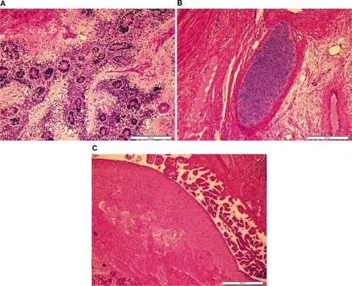
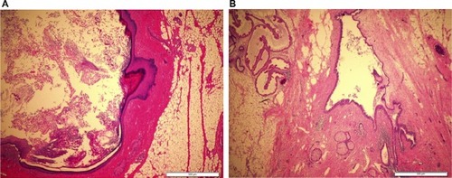
Figure 3 Histopathology of the recurrent tumor revealed heterogeneous mature elements including (A) epidermal cyst lined by mature keratinized squamous epithelium and filled with keratinous debris (H&E, 4×), (B) dermoid cyst lined by benign squamous epithelium surrounded by mature fibroadipose tissue with embedded adnexal glands (H&E, 4×).