Figures & data
Figure 1 Day 3 marked swelling, erythematous tense right eyelid presented at the first presentation.
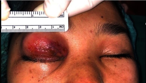
Figure 2 Day 3 (A) axial, (B) coronal CT scan of brain including orbit shows extensive right orbital inflammation with abscess formation, which extended medially from extraconal into intraconal space posterior to eyeball (arrow head).
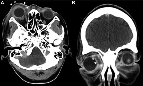
Figure 3 Day 3 (A) coronal and (B) axial T1W with fat suppression MRI scan of brain including orbit showed multiple hyposignal intensity areas with rim-enhancement in preseptal and intraconal spaces. The right superior ophthalmic vein thrombosis is suspicious by intraluminal filling defect with rim-enhancement (star) showing in (C and D) coronal view.
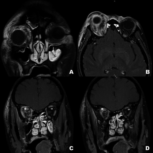
Figure 4 Day 7 limitation of extraocular movement of the right eye in (A) primary position, (B) left gaze, (C) right gaze, (D) up gaze, and (E) down gaze are demonstrated.

Figure 5 Color fundus photographs show (A) day 5 retinal whitening with scattered flame-shaped retinal hemorrhage, attenuated retinal arteries and venous dilatation representing combined central retinal artery and vein occlusion. There is optic disc swelling. The cilioretinal artery presenting with retinal whitening along the vessel demonstrates cilioretinal artery occlusion. (B) Day 10 a week after treatment by canthotomy and cantholysis to relieve orbital pressure, optic disc swelling, retinal swelling and whitening decreases. However, there is obvious evidence of a macular hole.
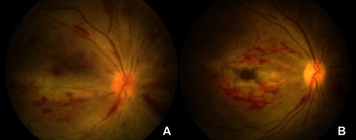
Figure 6 Day 10 fundus photograph shows (A) retinal whitening and hemorrhage in the macular area with macular hole. Cilioretinal artery is occluded (star). (B) Late phase of fluorescein angiography overlying on the fundus photograph reveals vascular filling defect of the arteries around the macula including cilioretinal branch (star).
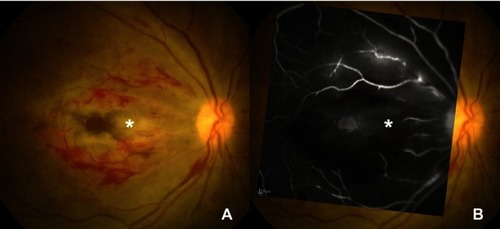
Figure 7 Day 10 fluorescein angiography in the early phase, (A and B) show delayed arterial filling, taken 10 seconds apart. (C) Vascular filling defect and perivascular leakage are detected in late phase with window defect of macular hole. Optical coherence tomography (OCT) corresponding to the upper section on (C) angiography depict (D) the inner retina thickening. This represents inner retinal swelling due to ischemia causing by arterial occlusion. (E) OCT passing the macular on (C) angiography reveals macular hole with the present of posterior hyaloid membrane. Thin strands connecting vitreous and retinal surface are demonstrated, confirming that the macular hole might be from ischemic process rather than traction force.
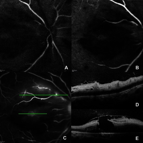
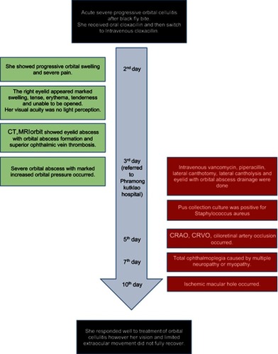
Figure 8 Timeline of case.