Figures & data
Figure 1 Chest radiograph demonstrating enlarged central pulmonary arteries and diminished peripheral pulmonary vascular marking.
Abbreviations: AP, anterior-posterior; R, right.
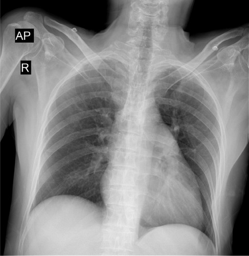
Figure 2 Transthoracic echocardiogram showing (left) right heart dilatation and (right) pulmonary hypertension.
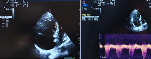
Figure 3 Pulmonary CT angiography demonstrates bilateral saddle pulmonary thromboemboli with right ventricle dilatation.
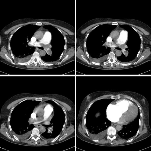
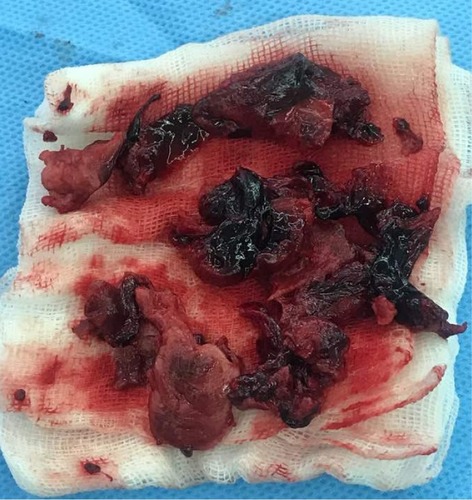
Figure 4 Thrombuses removed from the left and right pulmonary arteries at the time of surgery.