Figures & data
Figure 1 Main lesion with positive positron emission tomography–computed tomography (PET-CT).
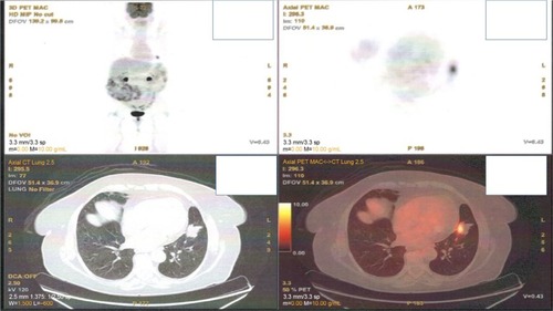
Figure 2 Positron emission tomography–computed tomography with lymph node station number 11 left (arrow).
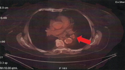
Figure 3 Left, computed tomography of the thorax with lymph node station number 11 left; middle, the carina of the left upper and lower lobe; and right, the lymph node 22 left with the convex probe ultrasound.
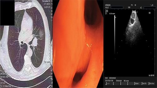
Figure 4 ALK gene rearrangement by FISH in the case of lung adenocarcinoma with positivity in 31% of neoplastic cells. FISH, ZytoLight SPEC ALK dual color, break apart probe (ZytoVision GmbH).
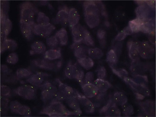
Figure 5 Positron emission tomography–computed tomography with the disease relapse lesion under the diaphragm 1.2 cm.
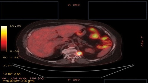
Figure 6 PD-L1 expression to the relapse site (nodule under the diaphragm).
Notes: (A) Programmed death-ligand 1-positive case with whole membranous staining (intense 1+ and 2+) in 90% of neoplastic cells; (B) hematoxylin/eosin of the same case (undifferentiated lung adenocarcinoma), magnification ×100.
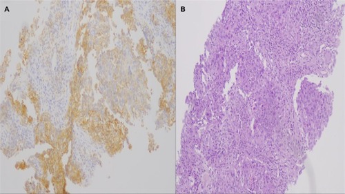
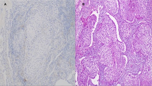
Figure 7 Main tumor PD-L1 expression after surgery.
Notes: (A) Programmed death-ligand 1-negative case without any membranous, whole or partially positive, magnification ×100; (B) hematoxylin/eosin of the same case with numerous neoplastic adenocarcinoma cells, magnification ×100.