Figures & data
Figure 1 (A) Major ulcers, 15 cm in diameter, with clear lines, elevated cyanotic edges on the front surface of the scrotum. The surface was presented by tender granulations, with parts showing yellow purulent discharge thickenings. On the internal surface of the hips there were ulcers 3×5 cm in size, with clear lines, even edges, and tender granulations on the bottom of the ulcers and yellow-green purulent discharge thickenings. The penis was deformed, and was actually presented by the urethra, covered with granulation tissue (before the treatment). (B) Occlusive dressing with an epithelizing ointment. (C) Complete healing through granulation and scarring of ulcers on completion of the 24-week treatment.
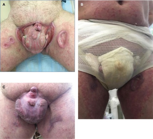
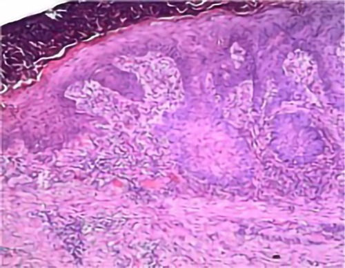
Figure 2 Lack of epidermis, infiltrate in the upper and mid dermis, which was mostly made up of leukocytes, lymphocytes, and histiocytes; absence of leukoplakia, widened vessel walls, gaping lumen, and disconnection between infiltrate and vessels (Magnification ×100).