Figures & data
Figure 1 The clinical manifestation of the patient with diagnosis of Buerger’s disease from early January 2018 until late May 2018.
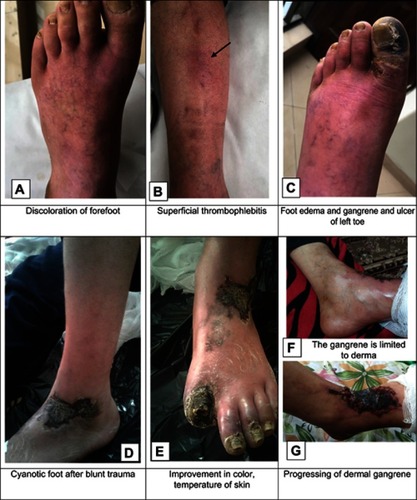
Figure 2 Computed tomography angiography of the patient with diagnosis of Buerger's disease. Occlusion of both superficial femoral arteries with multiple occlusions of left posterior tibial artery without stenosis in the aortoiliac and iliofemoral was observed.
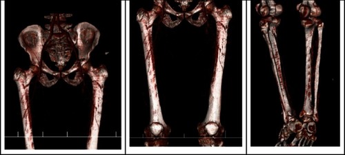
Figure 3 H&E staining; ×10 objective lens. The biopsy obtained around the ulcer of the amputated limb of a patient with diagnosis of Buerger’s disease
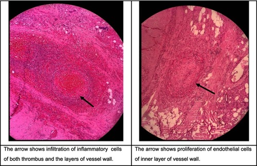
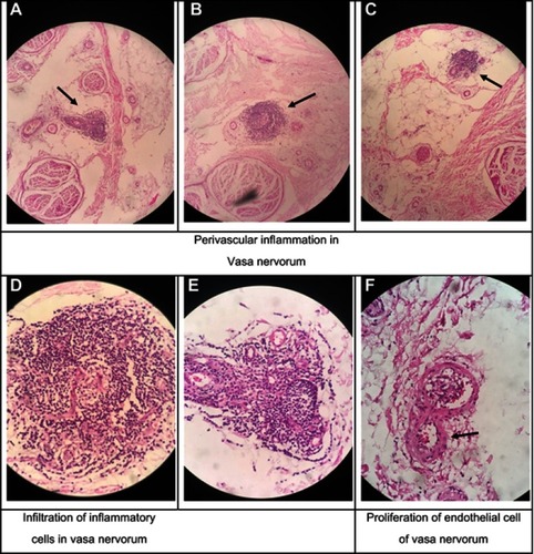
Figure 4 The biopsy obtained from the sural nerve of the amputated limb of a patient with diagnosis of Buerger’s disease.
Notes: H&E statining, ×5 objective lens, perivascular inflammation in vasa nervorum (A-C); H&E staining, 40× objective lens, infiltration of inflammatory cells and in vasa nervorum and proliferation of endothelial cells of vasa nervorum (D-F)