Figures & data
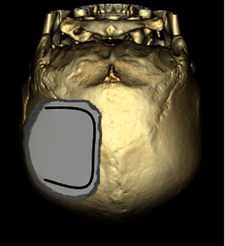
Figure 1 Serial CT brain imaging in follow-up, showing gradual enlargement of right tentorial SDH. (A) initial view of scanty right tentorial SDH, (B, C) status of SDH 16 and 26 hrs later, respectively, enlarging and propagating to right convexity, and (D) post-operative view.
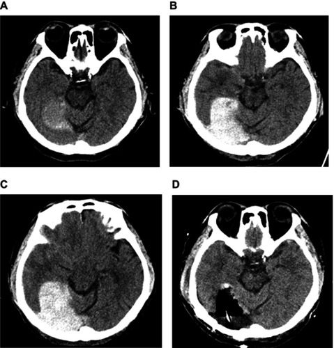
Figure 2 Pre- and post-operative CT images of the brain confirming evacuation of SDH and improvement of brain compression. (A, B) pre-, and (C, D) post-operative images (sagittal and coronal view, respectively).
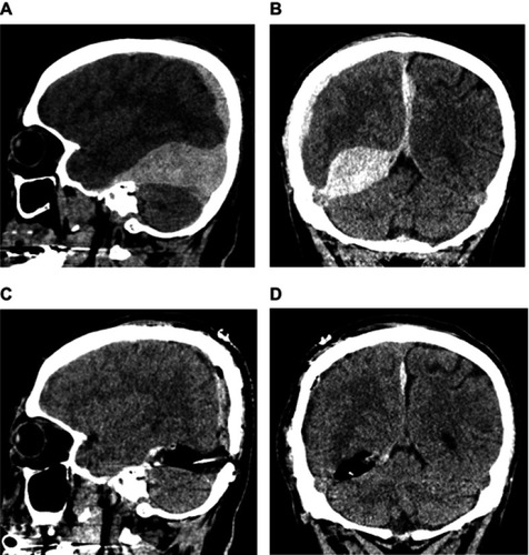
Figure 3 Illustration of the surgical approach depicting craniotomy and ⊃-shaped dural incision. The craniotomy site was approximately 7.5 cm×7.5 cm square. The distance from the medial margin of the craniotomy to the midline was 1.5 cm, and the distance from the lower margin to the transverse sinus was 1 cm.