Figures & data
Figure 1 In 2017, a 65-year-old female patient presented with left breast lumpiness to the symptomatic breast unit. Mammography revealed an indeterminate, low-density area within the upper outer quadrant of the left breast (long arrow). This was subject to core biopsies and subsequent titanium clip placement (short arrows).
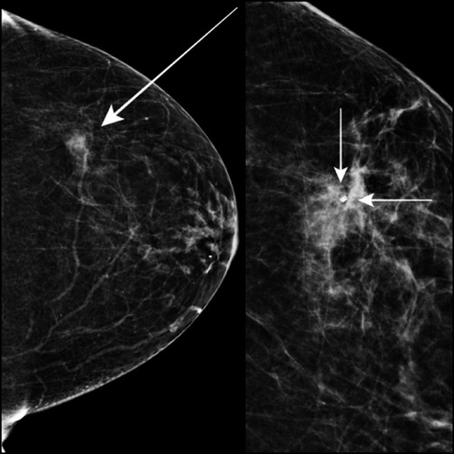
Figure 2 Seven weeks later, the same patient re-presented to the breast unit with a history of increasing pain and discomfort with regard to the marked upper outer left breast. High-resolution ultrasound revealed unremarkable titanium clip marker (arrow). There was no drainable collection or any significant findings to justify the symptoms. Location of the pain correlated well with the patient’s tenderness.
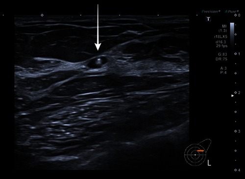
Figure 3 The same symptomatic patient requested removal of the metallic component and a vacuum clip excision was made. The procedure was successful, and titanium clip within the biopsy specimens was visualized (arrow).
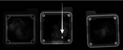
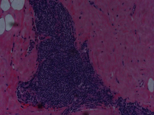
Figure 4 Microphotograph illustrates histopathology of the same patient as in –.