Figures & data

Figure 1 Black arrow showing a polycystic lesion in the right inguinal region along with serous fluid within, suggestive of hydrocele of the canal of Nuck.
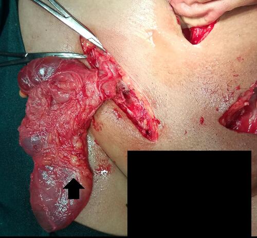
Figure 2 Incision sites in the right inguinal region, left inguinal region, and lower midline laparotomy. The black arrow pointing upward in the left inguinal area shows the omentum as a content of the left inguinal hernia; the downward arrow shows the site of the cystic component which ruptured during manipulation of the dilated canal of Nuck.
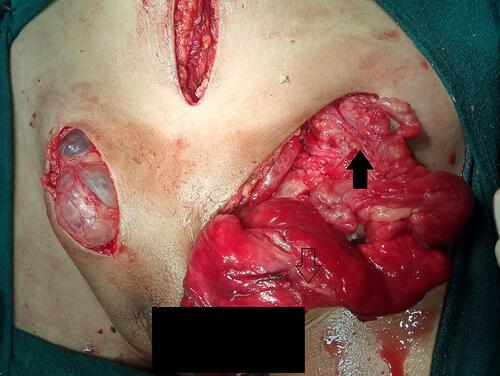
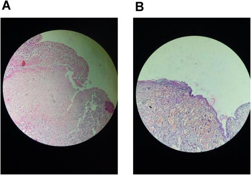
Figure 3 (A) Multiple sections examined from the bilateral inguinal region mass, with the cyst wall lined partly by flattened to cuboidal cells and partly by mesothelial cells, showing an area of necrosis with mixed inflammatory infiltrates. (B) Fibrocollagenous cyst wall lined by low cuboidal to flat epithelium along with proliferation of small-caliber vascular channels.