Figures & data
Figure 1 Clinical photograph. (A) Preoperative photograph showing proptosis of right eye. (B) Intraoperative photograph of right transcutaneous transeptal superior anterior orbitotomy and excisional biopsy. (C) One month postoperative photograph of right eye.
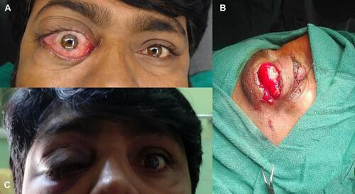
Figure 2 Clinical photograph. MRI T1, T2, FLAIR high signal intensity mass seen in intraconal compartment in lateral aspect of right orbit and MRI STIR images with lesion completely suppressed.
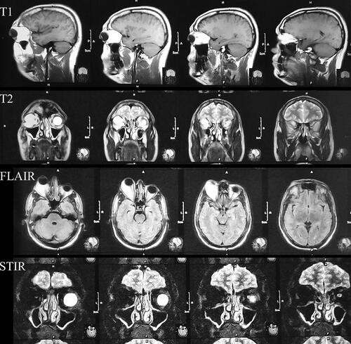
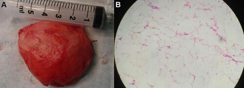
Figure 3 Clinical photograph. (A) Gross examination of specimen reveals a well encapsulated 4.3⨰4.5⨰2 cm, yellowish gray mass. (B) H&E stain shows tumor composed of lobules of mature adipocytes along with fibro collagenous septa (⨰10 magnification).