Figures & data
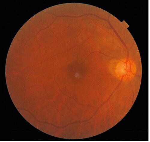
Figure 1 Right eye fundus photograph shows large cotton-wool spots in a peripapillary distribution as well as a heterogeneous pattern of grey translucency with periarterial sparing in the macula.
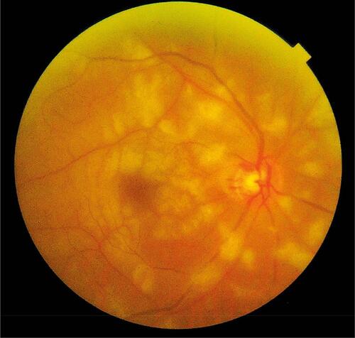
Figure 2 A horizontal spectral domain optical coherence tomography scan of the right eye was taken at foveal center, revealing an increase of the inner layer reflectivity and generalized inner retinal thickening.
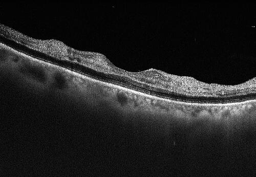
Figure 3 Right eye widefield fluorescein angiography shows delayed choroidal filling, taken 43 seconds after administering the dye.
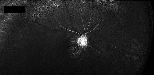
Figure 4 Right eye widefield fluorescein angiography shows peripheral pigmentary degeneration, discrete leakage and staining of the optic disc and peripapillary arterioles, taken 70 seconds after dye administration.
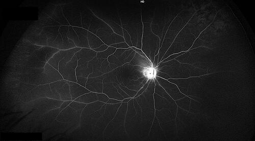
Figure 5 Right eye fundus photograph 4 weeks after initial presentation shows normal retinal characteristics.