Figures & data
Figure 1 White chorioretinal mass. Posterior uveitis with a white chorioretinal mass at the inferotemporal quadrant seen during the vitrectomy and the biopsy.
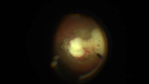
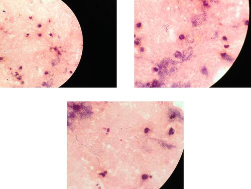
Figure 2 Left calf centesis. Disseminated Nocardia nova. Left calf centesis, 30 cc of pus. Direct examination with numerous polymorphonuclear neutrophils and aggregated gram positive rods and delicate, beaded, branching filaments. The Nocardia nova was found to be resistant to amoxicillin and clavulanate, tobramycin, ciprofloxacin and moxifloxacin.