Figures & data
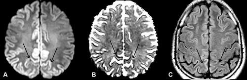
Figure 1 MRI brain on first admission. (A) (diffusion weighted imaging, DWI) and (B) (apparent diffusion coefficient, ADC): right > left restricted diffusion of parietal lobe (open arrows). (C) (fluid-attenuated inversion recovery, FLAIR): hyper-intense signal in right precuneus and parietal lobe (closed arrow), sparing white matter.
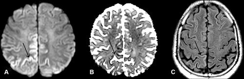
Table 1 Investigations Performed to Exclude Mimics and Chameleons of CJDCitation3
Figure 2 MRI brain on second admission one month later. (A) (DWI) (B) (ADC) and (C) (FLAIR): Bilateral symmetrical parietal cortical FLAIR hyperintense signals with restricted diffusion (open arrows) more marked in the parafalcine regions (closed arrow).