Figures & data
Figure 1 MRI scan sagittal and axial planes; (A–C) Sagittal T1WI demonstrated iso-intense intradural extramedullary lesion at different cuts from right to left; posterior scalloping of T11, (D) T2WI sagittal demonstrated heterogeneous mild hyper intensity of the lesion, (E) T2-STIR sagittal demonstrated homogenous marked hyper-intensity, (F) Post gadolinium sagittal demonstrated heterogeneous slight hyperintensity and well-enhanced rim (white arrow), G–J) Post enhanced axial demonstrated the extent of mass from T10/T11 disc (G and H) to T11/T12 disc (I and J). The mass lesion exited the left neural exit foramina of T11-T12 (black arrow, I). The lesion pulled the content of spinal canal towards itself (G and H). The well-enhanced rim was clearly demonstrated (white arrow, I).
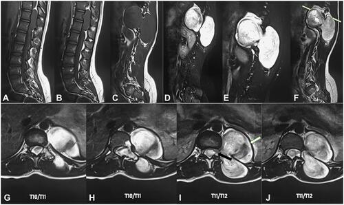
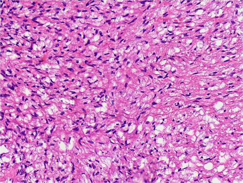
Figure 2 Hematoxylin and eosin showed spindle stromal cells, with many vessels and foamy vacuoles.