Figures & data
Table 1 Patient’s Characteristics
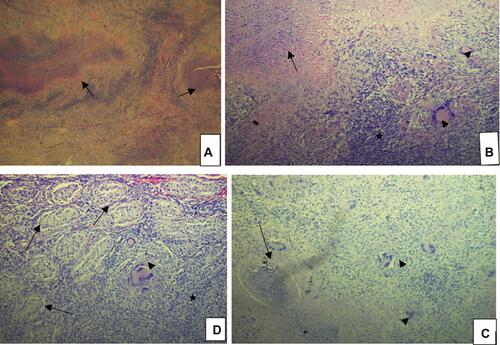
Figure 1 Normal chest X-ray. Histology of the left testis.
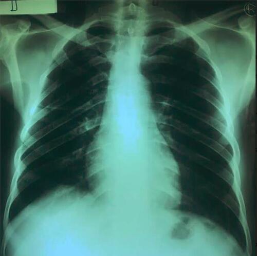
Figure 2 Shows the histology of the left testis (H&E stain). (A) Low power (×5); granulomas (arrows). (B) Low power (×10); caseous necrosis (arrow), numerous inflammatory cells (asterisk) and classic Langhan giant cells (arrowheads). (C) Low power (×10); numerous Langhan giant cells (arrowhead) and granuloma with caseation (arrow). (D) ×20 magnification showing atrophic seminiferous tubules (arrows) with interspersed inflammatory cells (asterisk) and Langhan giant cells (arrowhead).