Figures & data
Figure 1 Penetrating wound on left frontotemporal and preauricular region.
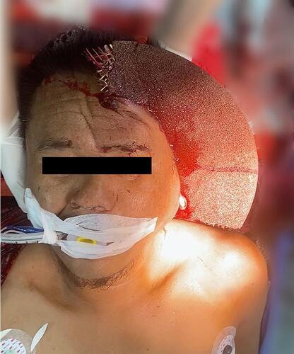
Figure 2 The transient left external carotid artery occlusion. (A) External carotid artery; (B) internal carotid artery; (C) common carotid artery.
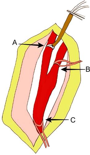
Figure 3 Technique of bleeding control. (A) External carotid artery occlusion; (B) internal carotid artery; (C) balloon tamponade; (D) common carotid artery.
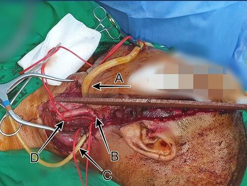
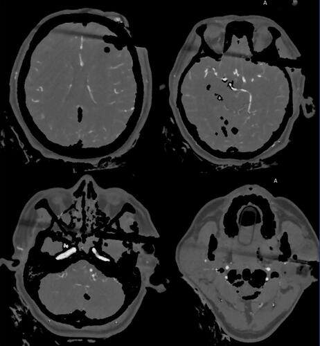
Figure 4 Computed tomography of a metallic foreign body with multiple left side skull and facial bone fracture.
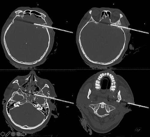
Figure 5 Computed tomography scan showing the presence of subarachnoid hemorrhage along left Sylvian fissure and acute subdural hemorrhage along frontotemporal convexity with brain swelling.
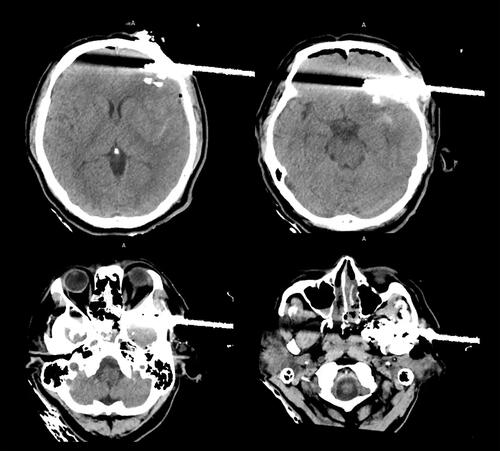
Figure 6 Computed tomography angiography shows no visible intracranial vascular injury.