Figures & data
Figure 1 (A and B) Fundus photograph of patient 1 with a bilateral moderate optic disc elevation. (C and D) Fundus fluorescein angiography revealed bilateral peripapillary microangiopathy and no optic nerve leakage. (E and F) Campimetric study showed cecocentral scotoma.
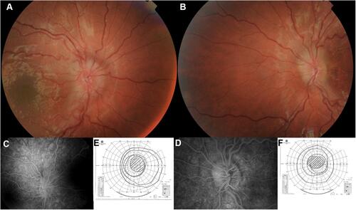
Figure 2 Brain and orbits magnetic resonance imaging (MIR) of patient 1. T2 hyperintensity in the posterior region of the left optic nerve and the optic chiasm.
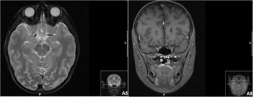
Figure 3 Spectral-domain optical coherence tomography (SD-OCT Spectralis, Heidelberg) analysis of patient 2. SD-OCT imaging documented peripapillary retinal nerve fiber layer (pRNFL) thickening in superior and inferior sectors.
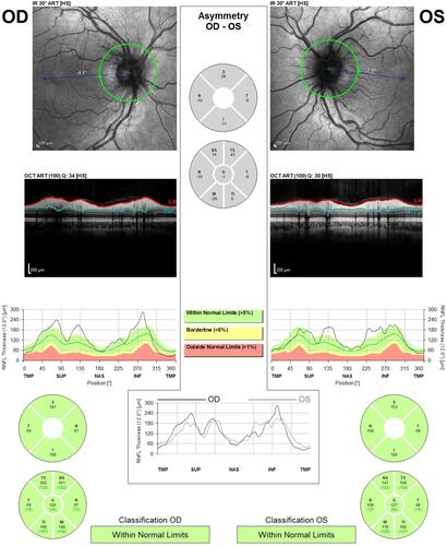
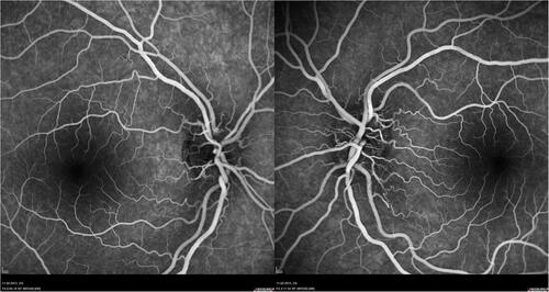
Figure 4 FA of patient 2 revealed vascular network papillary and peripapillary vascular microdilations, without diffusion in late stages.