Figures & data
Figure 1 (A and B) Fungating mass of approximately 15×15 cm noted at the left heel appreciated via lateral view (A) and inferior view (B).
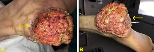
Figure 2 (A and B) Foot X-ray in anteroposterior view (A) and oblique view (B) showing lesion in the lateral posterior calcaneus with surrounding large irregular soft tissue radiopacity/mass. There was no bone resorption that may suggest osteomyelitis.
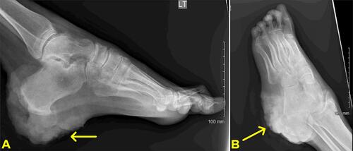
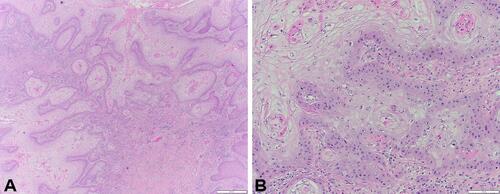
Figure 3 Photomicrograph of histopathologic specimen at low power reveals infiltration of tumor into deep dermis (A) and at high power (B) shows infiltrating islands of well-differentiated neoplasm and squamous epithelium within dermis.