Figures & data
Figure 1 Bilateral GPi-DBS in an adolescent DYT6 dystonia patient. (A) Image-guided frameless stereotaxic surgery was performed to implant DBS leads into GPi in the 12-year-old boy under general anesthesia. (B) Axial, (C) coronal and (D) sagittal images of O-arm (left panel, arrowheads), and fusion images with pre-operative MRI (right panel, arrows) indicating the location of DBS electrodes in bilateral GPi.
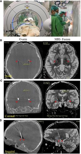
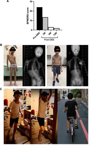
Figure 2 The clinical improvement of the DYT6 dystonia patient after effective bilateral pallidal stimulation. (A) The BFMDRS score changes at baseline and 3-, 6-, and 12-months after GPi-DBS. (B) Significant improvement in severe scoliosis of the patient and findings of spine x-rays pre-operatively and at 6-months after GPi-DBS. (C) Photos showing his daily physical activity nearly normal state after successful treatment of scoliosis and dystonia at 12 months after GPi-DBS.