Figures & data
Figure 1 Optical coherence tomography imaging of the patient’s right eye on initial presentation, demonstrating (A) an inferotemporal optic nerve pit with retinoschisis, intraretinal fluid, subretinal fluid, and (B) significant macular schisis.
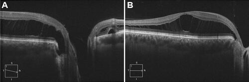
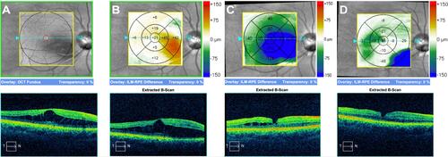
Figure 2 Optical coherence tomography macular change analysis showing structural evolution of the macula (A) at initial presentation, (B) 2 months post-operatively with persistence of macular schisis, (C) 10 months post-operatively with reduction of the intraretinal fluid following adjunctive use of oral spironolactone and topical dorzolamide and (D) 2 years post-operatively with almost complete resolution of the intraretinal fluid following long-term spironolactone use.