Figures & data
Figure 1 Arrows point to symblepharon formation seen inferiorly in the right (A) and left (B) eyes.
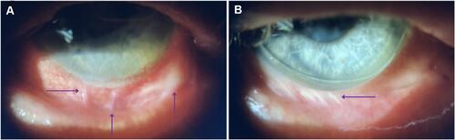
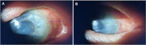
Figure 2 Limbal stem cell deficiency in the right (A) and left (B) eye. Arrows point to neovascularization and conjunctivalization of the cornea. Whorled keratopathy, obscured limbal architecture, subepithelial haze, and conjunctival hyperemia are also present bilaterally.