Figures & data
Figure 1 Ultrasound (US) images of a 25-year-old woman with a mature cystic teratoma. B-mode transabdominal US image showing a heterogeneous lesion with an echogenic mural nodule (dermoid plug) (red arrow).
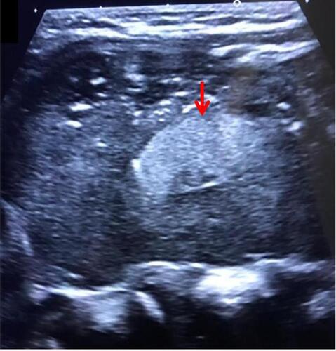
Figure 2 Axial contrast-enhanced abdominopelvic computed tomography images showing presence of enlarged uterus (red arrow) (A) consistent with early postpartum period and a normal appendix (red arrow) (B). Also note the non-enhancing ovarian mass (white arrow).
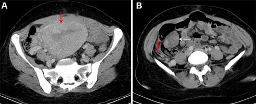
Figure 3 Axial contrast-enhanced abdominopelvic computed tomography image revealing intralesional fat (white arrow) and calcification (red arrow). Note the non-enhancing component of the lesion (arrowhead).
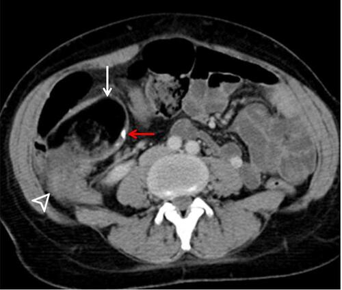
Figure 4 Surgical specimens showing unilocular cysts filled with sebaceous material, hair, and focal solid area with calcification area.
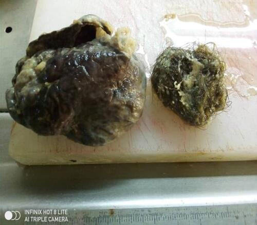
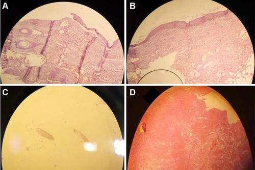
Figure 5 Low power microscopic view (A) showing tissue lined by stratified squamous epithelium with associated hair follicles and sebaceous glands. High power microscopic view (B) showing in the surface lined by stratified squamous epithelium with associated hair follicles and sebaceous glands. Microscopic (C); hair shaft material. Extensive hemorrhage, congestion, dilated blood vessels, and ischemia of mucosa and parenchyma (D).