Figures & data
Figure 1 Periorbital swelling of the right eye without erythema or pain.

Figure 2 The magnetic resonance imaging of the orbit. (A) Axial T2-weighted post-contrast images showed a mildly enhanced mass compressing the adjacent right lateral rectus muscle and the globe. (B) Coronal T2-weighted post-contrast images showed an extra-conal lesion, which was well-defined and iso-intense as the lateral rectus muscle.
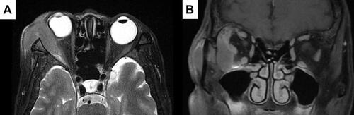
Figure 3 (A) Hematoxylin and eosin stain of the orbital mass revealed numerous blast cells with large round nuclei and prominent nucleoli. The immunohistochemical stain of the orbital mass showed positive expression of CD34 (B) and CD117 (C). (X400).
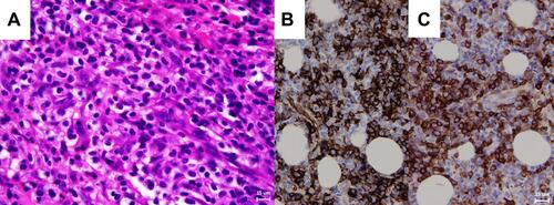
Figure 4 An allele-specific polymerase chain reaction disclosed the positive Val617Phe mutation of Janus Kinase 2 (JAK2) gene of the patient. (Lane 1).
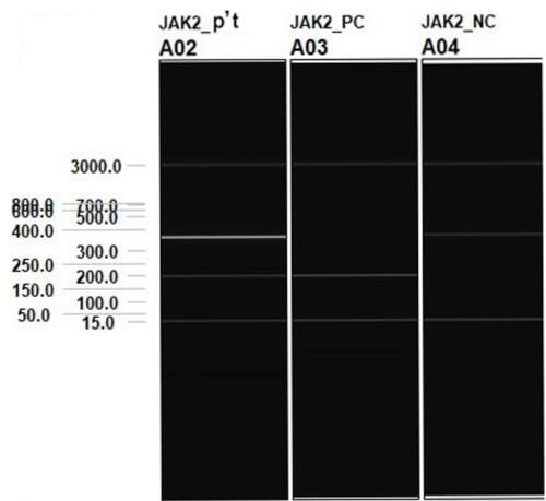
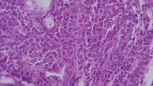
Figure 5 Marrow biopsy was done for increased blasts in peripheral blood and disclosed hypercellular marrow with around 80% cellularity. In HPF (X400) view, there was increased myeloid series and more than 20% blasts, indicating acute leukemia transformation.