Figures & data
Figure 1 Still picture of Supplementary Video S1 highlighting upper limb tremors and orofacial dyskinesias.
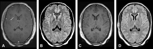
Figure 2 Axial magnetic resonance imaging (MRI) slice images of the brain: On admission: (A) T1-weighted contrast-enhanced (T1+C) and (B) axial fluid-attenuated inversion recovery (FLAIR) sequences illustrating contrast enhancement (white arrow) with sulcal hyper-intensity (black arrow) respectively; Day 15: (C) T1+C and (D) FLAIR sequences illustrating resolution of MRI abnormalities.