Figures & data
Figure 1 (A) scleral fixation using Yamane technique, (B) securing the haptics with cautery, (C) haptics trimming, (D) placing haptics in the scleral tunnel.
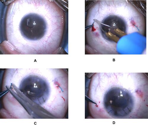
Figure 2 (A) scleral fixation using fibrin glue, (B) introducing IOL into the sulcus and externalizing the haptics through the flap, (C) haptics trimming, (D) scleral flaps and conjunctiva closed using fibrin glue.
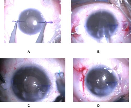
Figure 3 Postoperative anterior segment photos of the (A) right and (B) left eye.
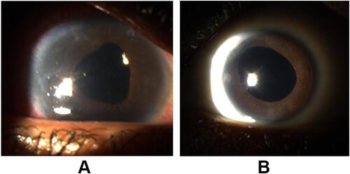
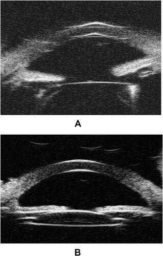
Figure 4 Ultrasound biomicroscopy of the (A) right and (B) left eye one year postoperatively.