Figures & data
Figure 1 3D-reconstruction (A and B) and lateral view in digital subtraction angiography (C) of left ICA presenting the fenestration with a flow-associated aneurysm within the proximal bifurcation. Vessel lumen diameters in mm as depicted in b: 1–2.77; 2–1.51; 3–1.90; 4–3.73; 5–3.46.
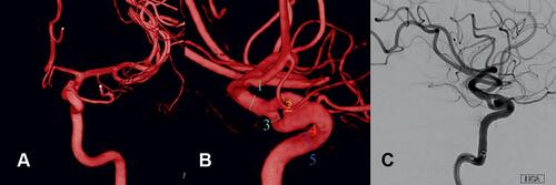
Figure 2 Angiographic follow-up via digital subtraction angiography of left ICA. (A and E) Directly after implantation of the pipeline device; (B and F) after three months; (C and G) after twelve months; (D and H) after twenty-four months. Note the transient relative hypoperfusion of the left anterior cerebral artery via left ICA three months post-interventionally that completely reversed during next follow-ups accompanied by transient intimal hyperplasia (white arrowhead).
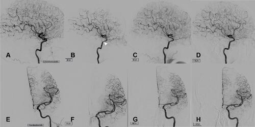
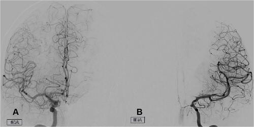
Figure 3 Coronal view of both ICAs via digital subtraction angiography three months after pipeline device implantation presenting left anterior cerebral artery hypoperfusion from left ICA (B) that is compensated via crossflow from right ICA (A).