Figures & data
Figure 1 The patient’s extraocular eye movements in different gaze positions. (A) The patient is looking straight showing no limitation of extraocular muscles. (B) The patient is looking to the left. There is no limitation of extraocular muscle movements. (C) The patient is looking to the right. There is limitation of abduction of the right eye indicating weakness of the right lateral rectus muscle due to sixth nerve palsy.
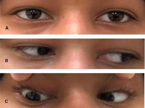
Figure 2 Average RNFL thickness is asymmetrical in both eyes with 58% of asymmetry. RNFL thickness values for different locations of the optic nerve scans showing the average thickness in μm in superior, temporal, inferior and nasal quadrants. The RNFL in the nasal quadrant of the right eye and left eye are 68 μm and 59 μm, respectively. Both readings are significantly thin compared with normal controls (96 μm). The RNFL in the temporal quadrant of the left eye is 55 μm which is severely thin compared with normal controls (73 μm). This thinning of RNFL quadrants is consistent with ON.
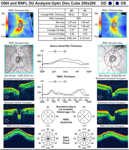
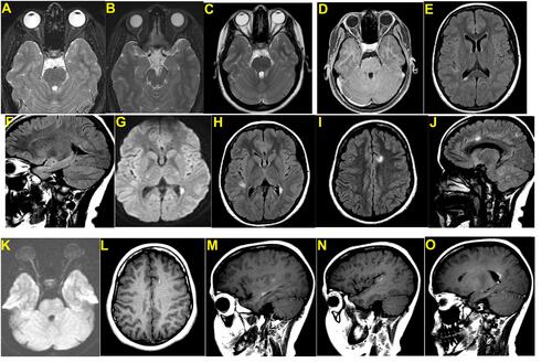
Figure 3 MRI of brain. (A) T2 fat sat image right optic nerve hyperintensity indicating optic neuritis. (B) T2 axial image showing left cerebral peduncle plaque. (C) T2 axial showing right optic neuritis. (D) T1 axial post-contrast image showing patchy enhancement of right optic nerve. (E) Periventricular T2 hyperintense focus in right peritrigonal region. (F) Flair sagittal image showing ependymal dot-dash sign sensitive in early MS. (G) Diffusion-weighted image showing left peri ventricular bright focus- T2 shine through effect. (H) Flair axial image showing right parietal juxtacortical and left periventricular foci. (I) Flair axial image showing left parafalcine frontal juxtacortical focus. (J) Sagittal flair image showing juxtacortical and infratentorial plaques. (K) Diffusion-weighted image showing bright right optic nerve. (L–N) Open ring enhancement of juxtacortical plaque. (O) Nodular enhancement in periventricular plaque.